Cotton Leaf Curl Virus is Distinct from Cotton Leaf Crumple Virus

Zhongguo Xiong, Athar Nadeem, Ziming Weng, and Merritt R. Nelson
Department of Plant Pathology, University of Arizona

Photographs and illustrations can be viewed by clicking the highlited texts. (Click on the picture to see the originals)


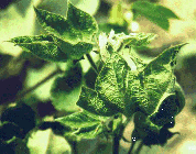
CLCurV symptoms

Fig. 1.

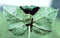
CLCurV symptoms

Fig. 2.

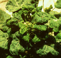
CLCV symptoms

Fig. 3.

Cotton leaf curl geminivirus (CLCuV) causes a major disease of cotton in Asia and Africa. Leaves of infected cotton curl upward (Fig. 1) and bear leaf-like enations on the underside along with vein thickening (Fig. 2). Plants infected early in the season are stunted and yield is reduced drastically. Severe epidemics of CLCuV have occurred in Pakistan in the past few years, with yield losses as high as 100% in fields where infection occurred early in the growing season.

Another cotton geminivirus, cotton leaf crumple virus (CLCrV), occurs in Arizona, California, and Mexico. CLCrV symptoms are distinguishable from CLCuV symptoms in that infected leaves curl downward accompanied by interveinal hypertrophy and foliar mosaic (Fig. 3).

Both CLCrV and CLCuV infect dicotelydonous plants and are whitefly-transmitted (Brown et al., 1983; Mansor et al., 1993). Previous studies (Brown and Nelson, 1984; 1987; Hameed et al., 1994; Mansor et al., 1993) suggested that they belong to the subgroup III geminiviruses. However, little information is available on the relationship of these two viruses with each other and with other subgroup III geminiviruses.

Fig. 4

In order to understand the nature and the relationship of this virus with other geminiviruses, particularly CLCrV, the DNA A of CLCuV was amplified by the polymerase chain reaction (PCR) as two fragments using primers specific for the DNA A of all whitefly transmitted geminiviruses. Two DNA fragments of about 1.2 kb and 1.5 kb were amplified (Fig. 4). In the same PCR primers, two DNA fragments of the CLCrV DNA A component were also amplified. These two fragments are approximately 1.2 kb and 1.4 kb, slightly smaller than the CLCuV DNA fragments. The difference in the size of DNA A components suggest that these two viruses are different.

Fig. 5

To further clarify the relationship between these two viruses, riboprobes complementary to the virion-sense strand of DNA were prepared using the cloned DNA A fragments. These probes were used to hybridized total cotton DNA extracted from CLCrV- and CLCuV-infected leaves. Under high stringency conditions, CLCrV and CLCuV probes only hybridized with DNA extracted from cotton tissues infected by the respective virus (Fig. 5). Neith probe hybridized with DNA extracted from leaf tissues infected by the other virus. Two DNA species corresponding to the single-stranded DNA encapsidated in geminivirus virions and the double-stranded replicative form (RF) were detected by this hybridization from each virus. In addition, a species of single-stranded DNA smaller than the genome-size was detected in CLCuV-infected tissue. This DNA presumably represents a defective subgenomic DNA of CLCuV. Similar subgenomic DNA has been reported in several other geminiviruses.
PCR-amplified DNA fragments from both CLCrV and CLCuV were subsequently sequenced. Analysis of nucleic acid sequences indicates that CLCuV is closely related to tomato yellow leaf curl virus, cassava latent virus, and Indian cassava mosaic virus from the old world, whereas CLCrV is more closely related to potato yellow mosaic virus, bean dwarf mosaic virus, and bean golden mosaic virus from the New World.

Together, these data clearly suggest that CLCrV occuring in North America and CLCuV occuring in Asia and Africa are two distinct viruses.

This page has been visited times.

iconPlease send your comments and suggestions to zxiong@ag.arizona.edu

Back to Xiong's Hompage